Figure 2.
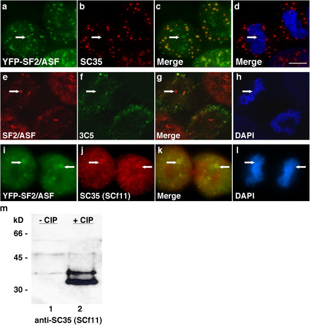
SR proteins in NAPs are hypophosphorylated. SC35 antibody (b, arrow), which recognizes hyperphosphorylated SC35, does not recognize the SC35 in NAPs (a, arrow) and is absent from the nucleus (c and d, arrows). 3C5 antibody that recognizes a family of hyperphosphorylated SR proteins (f, arrow) also does not recognize SR proteins in NAPs (e, arrow) and is absent from the nucleus (g and h, arrows). Projections of deconvolved image stacks illustrate the strictly cytoplasmic localization of hyperphosphorylated SR proteins as well as exclusion from nuclei (a–h). SCf11 (j, arrows) colocalizes with YFP-SF2/ASF in NAPs (i and k, arrows) and recognizes SC35 by immunoblot only when cell extract is treated with phosphatase (m, lane 2). Arrows in d, h, and l indicate NAP position. Bar, 5 μm.

