Figure 4.

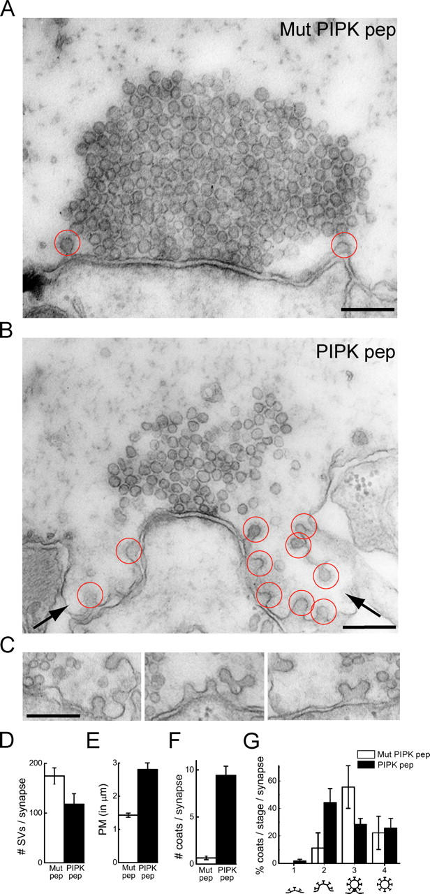
Ultrastructural changes produced by PIPK pep at lamprey synapses. (A and B) Electron micrographs of stimulated synapses after axonal injection of either Mut PIPK pep or PIPK pep. In the presence of the Mut PIPK pep (A), only few coated pits are observed. In contrast, numerous clathrin-coated pits (red circles) and large plasma membrane foldings (arrows) are observed in the presence of PIPK pep (B). (C) Gallery showing unconstricted clathrin-coated pits at periactive zones of synapses within PIPK pep–injected axons. Bars, 0.2 μm. (D–G) Quantification of the number of synaptic vesicles (D, SVs), the plasma membrane (PM) cross-sectional profile (E), the total number of clathrin-coated profiles (F), and the percentages of coated profiles at various stages of maturation (G) per synapse. Data represent mean values and SEM for 21 synapses from 2 axons injected with PIPK pep and 20 synapses from 2 axons injected with Mutant PIPK pep.
