
Figure 1.
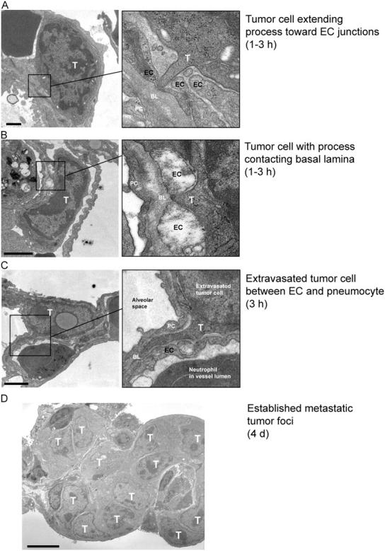
Tumor cell accumulation hours-to-days after tumor cell injection. To determine a time line for CT26 tumor cell extravasation after i.v. inoculation, we prepared lungs for analysis by transmission EM. By 1–3 h, tumor cells (T) had extended protrusions toward endothelial cells (EC) (A) or extended processes between EC junctions to contact the underlying basal lamina (BL) (B). (C) At 3 h, we observed individual extravasated tumor cells immediately outside the blood vessels where they were typically lodged in the extracellular space between an EC and a pneumocyte (PC). (D) This ultimately gave way to metastatic foci containing numerous tumor cells by day 4. Bars: (A–C) 1 μm; (D) 5 μm.