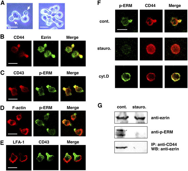
Figure 1.
EL4.G8 cells show polarized morphology with a clear uropod that is regulated by Ser/Thr kinases and the actin cytoskeleton. (A) Phase-contrast view showing the hand mirror–shaped morphology of EL4.G8 cells with clear uropods (left, arrows). Aggregation of EL4.G8 cells by their uropods (left). (B–E) Selective localization of the uropod markers (CD44 and CD43) and p-ERM in the EL4.G8 cell uropod, whereas F-actin and LFA-1 are enriched in the leading edge. Fixed, permeabilized cells were stained with fluorescence-labeled probes for cell surface CD44, CD43, or LFA-1, and for intracellular ezrin, p-ERM, or F-actin. (F) Staurosporine and cytochalasin D disrupt uropod structure. Cells were treated with DMSO (cont.), staurosporine (stauro.), or cytochalasin D (cyt.D) and stained for p-ERM and CD44. (G) Staurosporine abolishes ERM phosphorylation and the interaction between CD44 and ezrin in EL4.G8 cells. The total amount of ezrin and the p-ERM level were determined by Western blotting using lysates from control or staurosporine-treated cells (top and middle). CD44 ezrin interaction was detected by the immunoprecipitation of CD44 and Western blotting for ezrin (bottom). Bars (A–F), 10 μm.

