Figure 1.

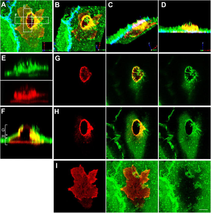
Monocytes migrate across the endothelium via transcellular routes in association with ICAM-1 projections. TNF-α–activated HUVECs were pretreated with MCP-1 and incubated with freshly isolated monocytes for 20 min. Cells were fixed and stained for ICAM-1 (CBRIC1/11-488; green), LFA-1 (CBR-LFA1/7-Cy3; red), and VE-cadherin (55-7H1-Cy5; blue; shown in A–D only) and imaged by confocal microscopy. (A) Top view projection of all z-series sections of a representative monocyte transmigrating via a transcellular route. (B–D) Field in A was rendered as a series of three-dimensional projections, each representing successive rotation about both the x and z axis in 30° intervals for a total of 90° about each axis. (E) Side view projection of cross section E in A depicting ICAM-1 projections (top) and linear LFA-1 clusters (bottom) separately. (F) Side view projection of cross section F in A. Apical (G), middle (H), or basal (I) z-axis sections, as indicated by brackets in F, are projected as top views. Bar, 5 μm.
