Figure 2.
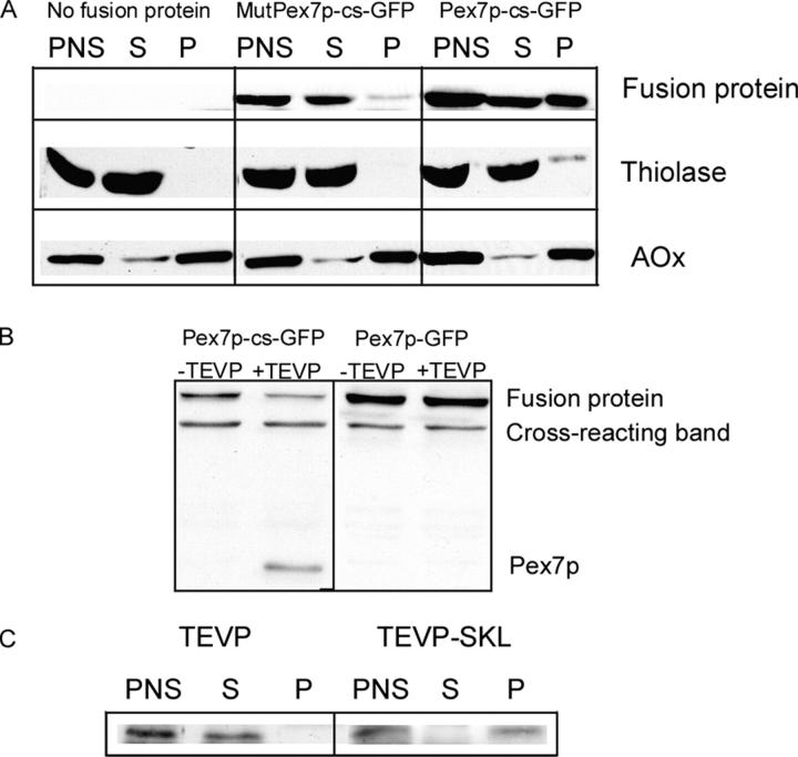
Intracellular distribution and cleavability of Pex7p-cs-GFP. (A) Cell fractionation of Δpex7 cells expressing Pex7p-cs-GFP (right) or MutPex7p-cs-GFP (middle) or no fusion protein (left). Immunoblots with anti-Pex7p, anti-thiolase, and anti-AOx antibodies. (B) In vitro cleavage of the fusion protein. Total cell lysates of Δpex7 cells, expressing either Pex7p-cs-GFP or Pex7p-GFP, were treated with or without purified TEVP (Invitrogen) for 30 min at 30°C. Pex7p cleavage product is seen only in the second lane that has Pex7p-cs-GFP treated with TEVP. (C) Intracellular distribution of TEVP and TEVP-SKL in Δpex7 cells. Immunoblot with anti-TEVP.

