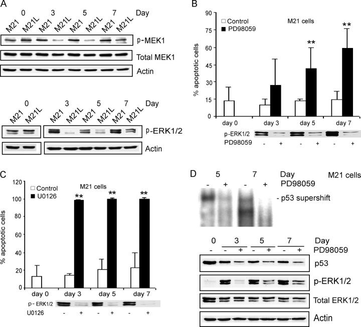
Figure 5.
Integrin αv-dependent MEK1 activity is required for melanoma cell survival in 3D-collagen. (A) M21 (αv+) cells and M21L (αv−) cells were cultured under 2D conditions (d 0) and within 3D-collagen for the indicated times. The levels of active MEK1 and ERK1/2 as well as of total MEK1 and ERK1/2 were detected by Western blotting, with actin as control and the displayed blots are representative among five experiments. (B and C) M21 (αv+) cells within 3D-collagen were treated with the MEK1 inhibitors PD98059 (B) and U0126 (C) or DMSO as a vehicle control. Annexin-V staining detected apoptosis and the bar graphs show the mean ± SD of apoptotic cells among three independent experiments (** −P < 0.01, as compared with vehicle control using unpaired two-tailed t test). Phosphorylated ERK1/2 was detected by Western blotting to control for the suppressive effect of the MEK1 inhibitors. (D) p53 DNA-binding activities were detected by EMSA and p53 protein levels determined by Western blotting in M21 (αv+) cells treated with PD98059 within 3D-collagen for 3–7 d. Phosphorylated ERK1/2 was detected to monitor the inhibitory effect of PD98059, and total ERK1/2 and actin levels were analyzed as controls. Note that the exposure time in this EMSA was longer than for EMSAs displayed in other figures (Fig. 2, B and D).

