Figure 1.
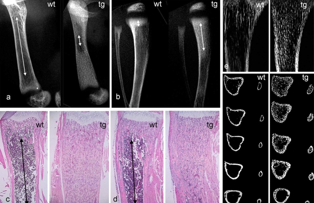
Impaired development of the marrow cavity and hematopoietic tissue in long bones of COL1-caPPR mice. (a) High resolution radiograms of the femurs of wt and tg mice at 2 wk. Note the marked difference in length of the marrow cavity (arrows). (b) High resolution radiograms of tibiae and fibulae of wt and tg mice at 2 wk. Note the different lengths of the primary spongiosa (arrows). (c and d) Histological sections of the distal metaphysis of the femur (c) and proximal metaphysis of the tibia (d) at 2 wk. Red marrow extends to the metaphyseal end of the primary spongiosa in wt mice (arrows), but only medullary bone is present in tg mice. (e and f) High resolution contact microradiography (e) and microCT analysis (f) of tibiae at 2 wk. The excess medullary bone formed in tg mice is obvious with both techniques. MicroCT demonstrates that the normal partition between cortical bone and marrow space is lost in tg mice, and that both are replaced by a continuous plexus of cancellous bone. Sections extend from 0.5 to 2.5 mm below the physis.

