
Figure 2.
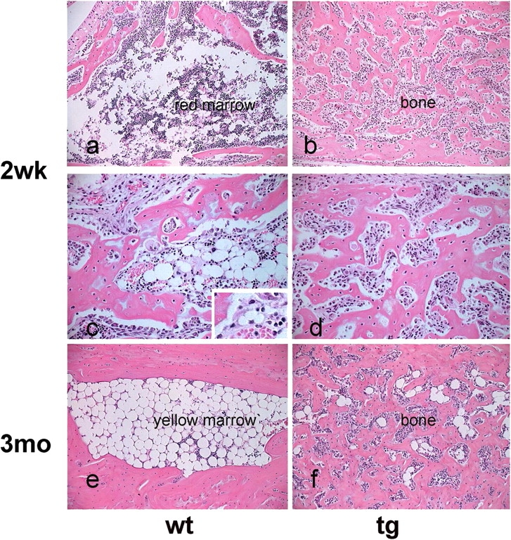
Impaired development of adipose (yellow) marrow in COL1-caPPR mice. Histology of the calcaneum at 2 wk and 3 mo. At 2 wk, the wt marrow cavity contains red hematopoietic marrow (a) with scattered adipocytes, and frequent multivacuolar, developing adipocytes (c and inset). In tg mice, a distinct cavity is not observed, hematopoiesis and adipocytes are absent, and an excess of bone is present (b and d). At 3 mo, hematopoiesis is no longer present, and the marrow cavity is filled with mature adipocytes in wt mice (e). No cavity is present in tg mice, and only rare adipocytes are found in narrow vascular spaces interrupting the continuity of the excess bone (f).