
Figure 5.
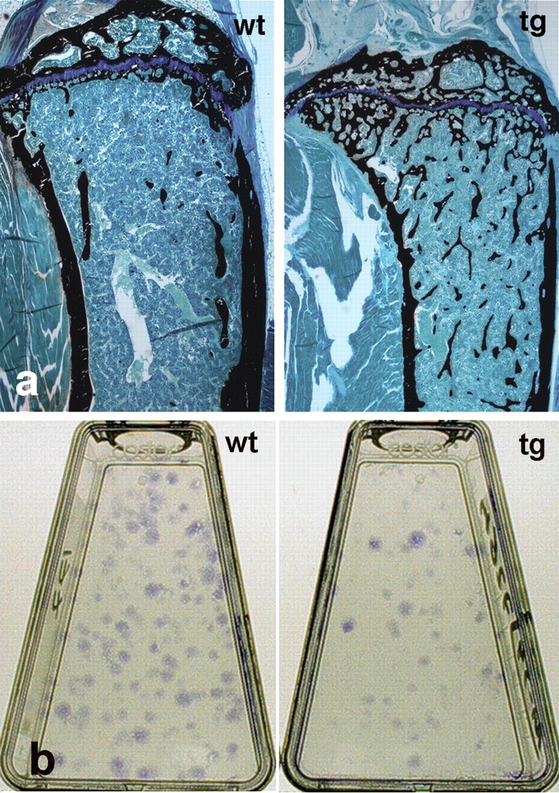
Histology of the bone/marrow organ and CFU-F frequency in mice at skeletal maturity. (a) Histology of the proximal metaphysis of the tibia in wt and tg mice at 4.5 mo. Hematopoietic marrow now fills the marrow cavity up to the physis in both wt and tg mice. A marked excess of trabecular bone is observed in tg mice compared with in wt mice (undecalcified MMA sections, von Kossa staining). (b) Representative primary cultures of cells established at clonal density from wt and tg mice. Note the higher number of colonies (CFU-F) in wt cultures.