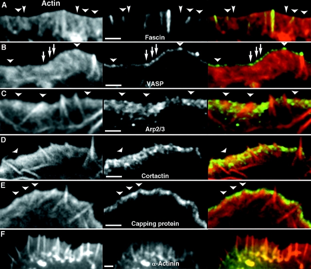
Figure 4.
Localization of filopodial and lamellipodial markers in Λ-precursors. Left column; actin revealed by Texas Red-phalloidin (A, B, and F) or by GFP-actin expression (C–E). Central column; actin-binding proteins (as indicated) revealed by expression of GFP-fusion proteins (A, B, and F) or by immunostaining (C–E). Right column; merged images. Λ-precursors are indicated by arrowheads. (A) Fascin is strongly enriched in established filopodia and localizes to the distal section of some Λ-precursors (wide arrowheads), but not others (narrow arrowheads). (B) VASP forms bright dots at the tips of filopodia and Λ-precursors. Additional dots can be seen along the leading edge (arrows), which apparently do not correspond to Λ-precursors. (C–E) Lamellipodial markers, Arp2/3 complex (C), cortactin (D), and capping protein (E), are excluded from filopodia and are partially depleted from Λ-precursors. (F) α-Actinin localizes to proximal parts of lamellipodia and filopodia. Bars, 2 μm.

