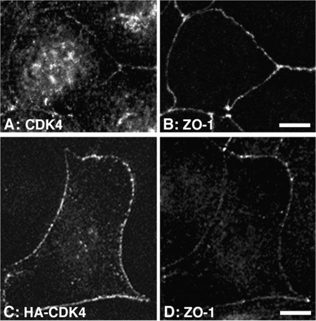
Figure 7.
Association of CDK4 with intercellular junctions. (A and B) MDCK cells were plated on coverslips and grown for 3 d. The cells were then fixed with methanol and processed for double immunofluorescence using rabbit anti-CDK4 and rat anti–ZO-1 antibodies. Shown are confocal XY-sections (A, CDK4; B, ZO-1 staining). (C and D) Confocal XY-sections of MDCK cells transiently transfected with a cDNA coding for HA-tagged CDK4. The cells were stained with rabbit anti-HA and rat anti–ZO-1 antibodies (C, HA-CDK4; D, ZO-1 staining). Bars, 10 μm.

