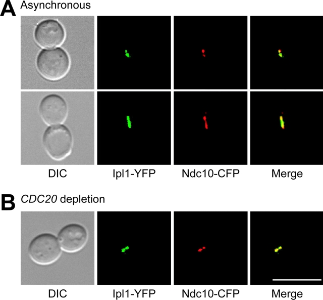
Figure 2.
Ipl1p localizes to metaphase kinetochores that are under tension. (A) Microscopy was performed on cells containing Ipl1–YFP (green) and Ndc10–CFP (red) (SBY1246). DIC pictures are shown on the far left. The merged image (yellow, far right) shows that Ipl1p and Ndc10p colocalize in metaphase cells where kinetochores are precociously separated (top). Some cells also show Ipl1p and Ndc10p costaining on the short spindle (bottom). (B) pGAL-CDC20 cells containing Ipl1–YFP and Ndc10–CFP (SBY1246) were arrested in metaphase by shifting the cells to glucose medium. The merged microscopy images show that Ipl1p and Ndc10p also colocalize in metaphase-arrested cells. Bar, 10 μm.

