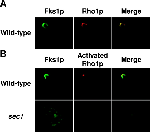
Figure 5.
Distribution of wild-type Rho1p and activated Rho1p. (A) Colocalization of wild-type Rho1p and Fks1p/2p. Wild-type cells were cultured at 25°C, fixed, and stained for immunofluorescence microscopy with the anti-Fks1p/2p antibody (green) or the anti-Rho1p antibody (red). (B) Localization of activated Rho1p to a restricted region on the plasma membrane in wild-type cells. Wild-type cells were cultured at 25°C (top panels), whereas sec1 mutant cells were incubated at 37°C for 2 h (bottom panels), and stained for immunofluorescence microscopy with the anti-Fks1p/2p antibody (green) or the anti-actRho1p antibody (red).

