
Figure 8.
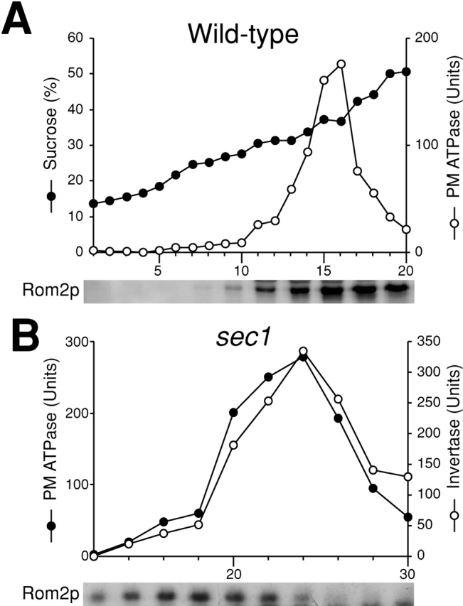
Subcellular fractionation analysis of Rom2p when vesicular transport is blocked. (A) Distribution of Rom2p on the plasma membrane in wild-type cells. Wild-type cells were incubated in YPD at 37°C for 2 h. Cell lysate was fractionated on a 20–60% sucrose density gradient. Top, distribution of plasma membrane ATPase (open circles) and sucrose density (closed circles). Bottom, immunoblotting analysis of Rom2-HA with an anti-HA antibody. (B) Distribution of Rom2p in Sephacryl™ S-1000 column fractions of sec1 cells. Top, distribution of plasma membrane ATPase (closed circles) and invertase (open circles). Bottom, immunoblotting analysis of Rom2-HA.