
Figure 2.
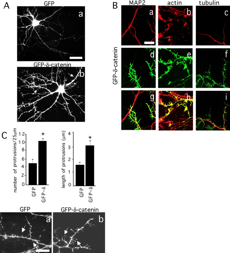
δ-Catenin induces protrusions along the distal dendritic shafts of hippocampal neurons. (A) Rat hippocampal neurons in culture for 8 d were transfected with GFP (a) and GFP–δ-catenin constructs (b) and fixed 24 h later. Small branches and filopodial-like protrusions on dendrites are indicated by arrows in b. Bar, 15 μm. (B) Hippocampal neurons in culture for 8 d were transfected with GFP–δ-catenin (d–f) and stained for MAP2 (a), actin (b), and tubulin (c). Panels g–i show colocalization. Bar, 5 μm. (C) Quantification of the number and length of protrusions for GFP and GFP–δ-catenin–transfected neurons. Detail of the protrusions is shown for GFP- (a) and GFP–δ-catenin– (b) transfected neurons. Bar, 5 μm.