
Figure 6.
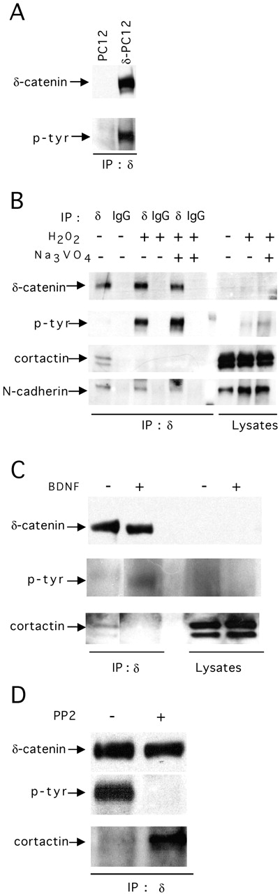
δ-Catenin–cortactin interaction is regulated by tyrosine phosphorylation. (A) PC12 and δ-PC12 cells were lysed and immunoprecipitated with mAb δ-catenin. Blots were immunolabeled with a mAb δ-catenin or a phosphotyrosine antibody (p-tyr). (B) Hippocampal neurons in culture for 10 d were treated for 5 min with 200 μM H2O2 or 200 μM H2O2 plus 1 mM sodium orthovanadate. Cells were lysed and immunoprecipitated by a mAb δ-catenin. Blots were immunolabeled for mAb δ-catenin, phosphotyrosine, cortactin, and N-cadherin. (C) Hippocampal neurons in culture for 3 wk were treated with BDNF (50 ng/ml) for 1 h, and δ-catenin was immunoprecipitated with a mAb δ-catenin. Blots of the immunoprecipitated products were labeled with mAb δ-catenin, phosphotyrosine, and cortactin antibodies. (D) δ-PC12 cells (pretreated with 100 ng/ml NGF for 36 h) were treated with PP2 (25 μM for 1 h). Immunoprecipitated products were blotted with mAb δ-catenin, phosphotyrosine, and cortactin antibodies.