
Figure 7.
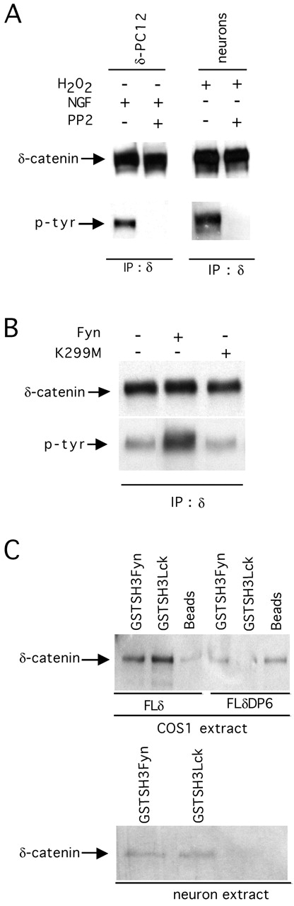
Tyrosine phosphorylation of δ-catenin. (A) δ-PC12 cells (pretreated with NGF for 36 h) and hippocampal neurons were treated with 25 μm PP2 for 1 h; additionally, hippocampal neurons were treated with 200 μM H2O2 for 5 min, δ-catenin was immuno- precipitated with a mAb δ-catenin, and its phosphorylation state was tested with antiphosphotyrosine. (B) δ-PC12 cells were transfected with constructs corresponding to Fyn or a dominant-negative Fyn (FynK299M). Cells were lysed, and lysates were subjected to immunoprecipitation with a mAb δ-catenin. The immunocomplexes were sequentially immunoblotted with a mAb δ-catenin and a phospho- tyrosine antibody. (C) In the top panel, COS 1 cells were transfected with FLδ or FLδΔP6. Cell lysates were incubated with GST–SH3Lck and GST–SH3Fyn fusion proteins bound to glutathione-sepharose beads. Lysates incubated with glutathione beads alone served as a control. In the bottom panel, GST–SH3Lck and GST–SH3Fyn fusion proteins bound to glutathione sepharose beads, or beads alone were incubated with lysates from 3-wk-old hippocampal neurons. In all lanes, eluted proteins were blotted with a mAb δ-catenin.