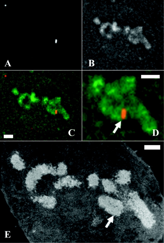
Figure 7.

Normal chromosome morphology over insert regions—correlative light and electron microscopy. In native chromosomes of clone Con1, vector inserts appear as a band going over the entire width of the chromosome. Sections are 0.2 μm thick. (A–C) Fluorescent light microscopy of a single section; lac repressor immunostaining signal, staining for total DNA with DAPI, combined A and B, respectively. (D) Two-fold expanded image from C. (E) An EM image of the same section. Arrows indicate insert region labeled with immunofluorescence probes (D) and corresponding regions on EM sections (E). Bars, 1 μm.
