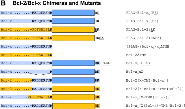
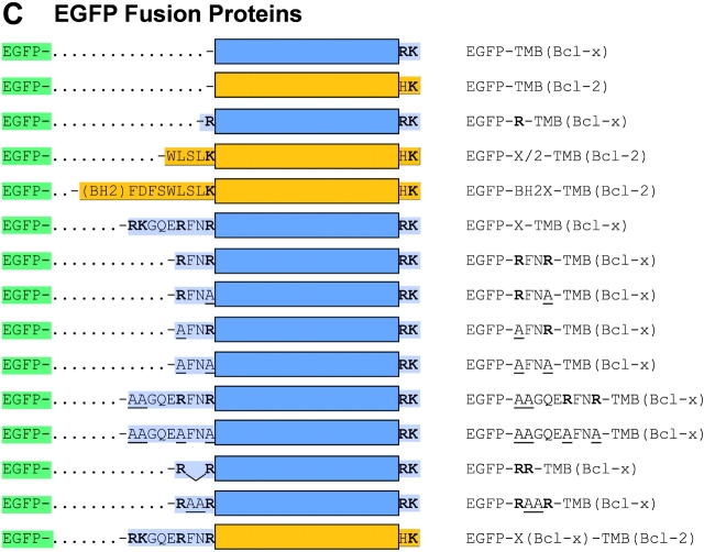
Figure 2.
Schematic representation of Bcl-2, Bcl-xL, Bcl-xS, their mutants, and the EGFP fusion constructs. Schematic structure and amino acid sequences of (A) the COOH-terminal parts of wild-type Bcl-2 (yellow) and Bcl-xL/xS (blue), including the 19–amino acid-long TM domain, flanked by one to two basic amino acids at one end (B) and the X or X/2 domain (half of the X domain) at the other end (basic amino acids are numbered and indicated in bold); (B) the COOH-terminal parts of Bcl-2 and Bcl-xL/xS mutants (point mutations and insertions are underlined); (C) the COOH-terminal mutants of Bcl-2 and Bcl-x fused to the COOH terminus of EGFP.


