
Figure 2.
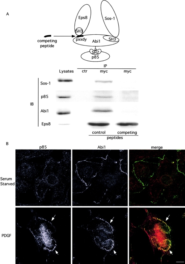
p85 associates with the Eps8–Abi1–Sos-1 complex under physiological conditions. (A) Top; model of the Eps8–Abi1–p85–Sos-1 complex. Bottom; lysates (10 mg) from −/− [Eps8myc] cells and supplemental materials) were immunoprecipitated (IP) with the indicated antibodies (ctr, irrelevant antibody) in the presence of the peptides PPPPPVDYTEDEE (competing) or PPPPPVAATEDEE (control). Immunoblotting (IB) was performed with the indicated antibodies. (B) Quiescent (Serum Starved) and PDGF-treated (PDGF) MEFs were fixed and stained with the indicated antibodies. Confocal analysis was carried and the apical sections are shown. Notably, colocalization between Abi1 and p85 could be detected only when the apical sections of PDGF-treated cells were analyzed. In these sections, the increase in the local concentration of the two proteins favors their detection by immunofluorescence, consistent with a notion that a relatively small (but physiologically relevant) pool of p85 is associated with Abi1 and enriched in ruffles. Arrows indicate dorsal ruffles. Bar, 10 μm.