
Figure 1.
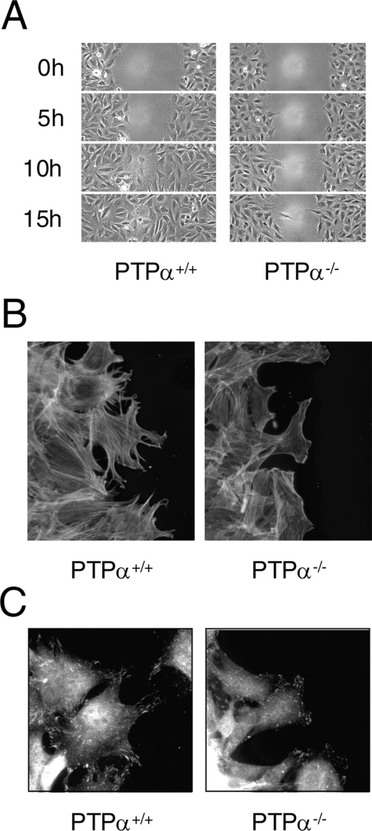
Defective migration of PTPα − / − cells. (A) Delayed migration of cells lacking PTPα. The migration of PTPα+/+ and PTPα−/− fibroblasts into an empty area of the culture dish was followed by time-lapse video microscopy. Fields at the indicated times are shown. (B) 6 h after wounding, cells at the migrating edge were stained for actin with Alexa Fluor 488–conjugated phalloidin. (C) Cells at the leading edge were stained with anti-phospho-Tyr397 FAK antibody just before wound closure of the PTPα+/+ cells.