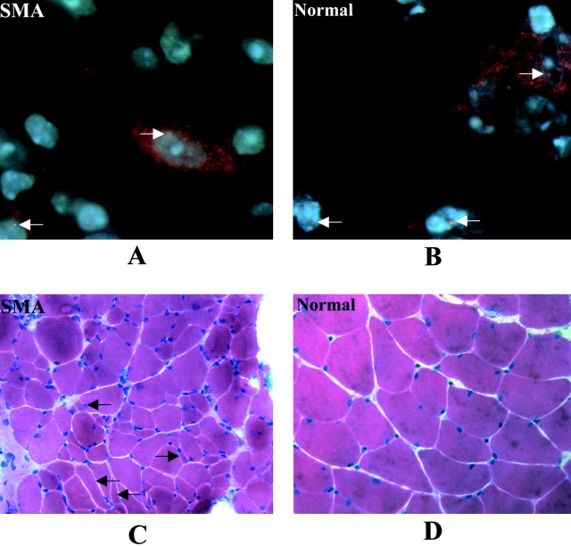
Figure 4.
Immunocytochemical staining of spinal cord, and hematoxylin and eosin staining of muscle. Staining of SMN in spinal cord sections of (A) 1-mo-old SMN A2G;SMN2;Smn − / − type III SMA mouse and (B) an age-matched Smn + / −control. Type III SMA mice express SMN in the motor neurons (arrows). However, nuclear gems in these animals are less intense and not as numerous as those seen in normal littermates. (600× magnification). Gastrocnemius from the same animals was sectioned and stained with hematoxylin and eosin. Numerous angulated and atrophied fibers (arrows) are evident in (C) type III SMA muscle as compared with (D) normal muscle (200× magnification).

