
Figure 1.
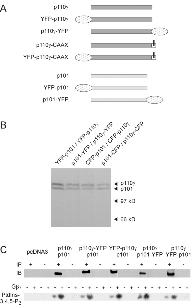
Construction and characterization of fluorescent PI3Kγ fusion proteins. (A) Schematic representation of the wild-type, fluorescent, and membrane-targeted PI3Kγ subunits. A YFP (or CFP) tag was fused to either the NH2- or the COOH terminus of p110γ and p101. The p110γ-CAAX fusion protein contains an isoprenylation motif (bars for lipid modifications). (B) Differently fluorescence-tagged PI3Kγ subunits were coexpressed in HEK cells, and cell lysates were subjected to SDS-PAGE followed by immunoblotting with a GFP-specific antibody. (C) Catalytic activity of fluorescent PI3Kγ fusion proteins. HEK cells were transfected with the indicated plasmids. Lysates were subjected to immunoprecipitation (IP) with (+) or without (−) an anti-p110γ antibody. immunoprecipitation was controlled by immunoblotting (IB) with another anti-p110γ antibody. Shown are chemiluminescence images (top panels). Immunoprecipitates were assayed for in vitro PI3K activity in the absence or presence of 120 nM Gβγ using PtdIns-4,5-P2– containing lipid vesicles and γ[32P]ATP as substrates. Depicted are autoradiographs of the generated 32P-PtdIns-3,4,5-P3 (bottom panels).