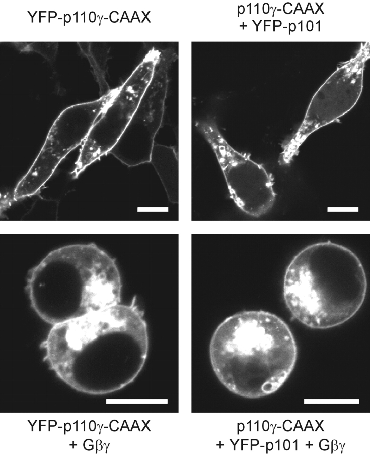
Figure 6.
Characterization of a constitutively membrane-associated p110γ-CAAX. HEK cells were transfected with the indicated plasmids and analyzed by confocal laser scanning microscopy. Images of typical cells are shown. White bars indicate a 10-μm scale. Top panel; Subcellular localization of YFP-p110γ-CAAX (left) or YFP-p101 expressed together with p110γ-CAAX (right). Bottom panel; Coexpression with Gβγ.

