
Figure 2.
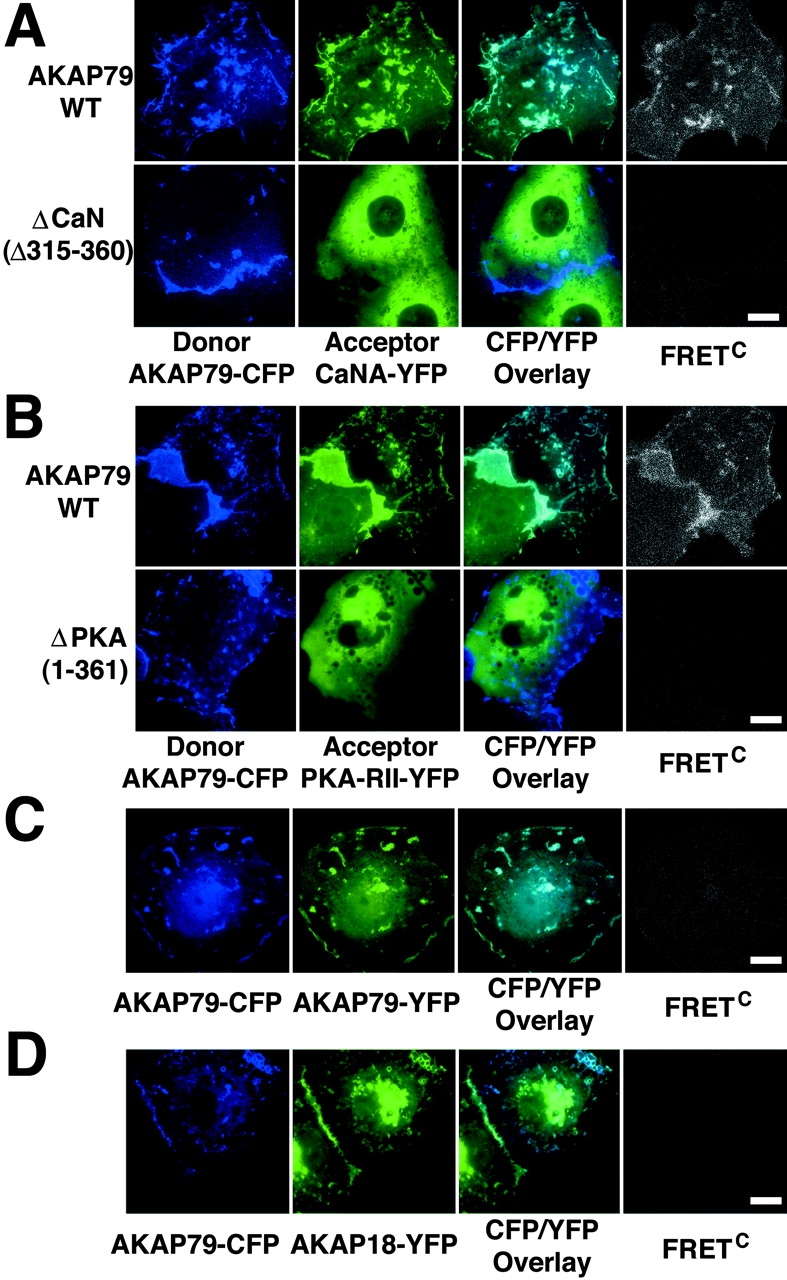
CFP/YFP micro-FRET microscopy imaging of AKAP79 binding to CaN and PKA in living cells. (A) Plasma membrane/cortical colocalization (CFP/YFP Overlay) and direct binding (FRETC) seen for CaNA–YFP (green) and AKAP79–CFP WT but not ΔCaN (Δ315–360) (blue) in live COS7 cells. (B) Plasma membrane/cortical colocalization (CFP/YFP Overlay) and direct binding (FRETC) seen for PKA-RIIα–YFP (green) and AKAP79–CFP WT but not ΔPKA (1–361) (blue) in live COS7 cells. (C) Negative control showing no FRETC for AKAP79–CFP (blue) and AKAP79–YFP (green) that colocalize at the plasma membrane (CFP/YFP Overlay) but do not bind to each other. (D) Negative control showing no FRETC for AKAP79–CFP (blue) and AKAP18(1–16)–YFP (green) that colocalize (CFP/YFP Overlay) but do not bind to each other. Bars, ∼15 μm.