Figure 4.
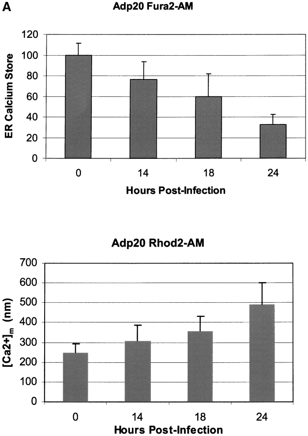
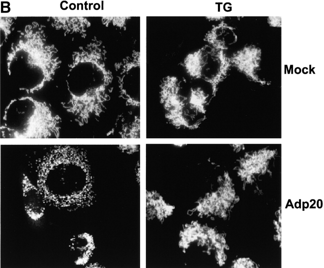
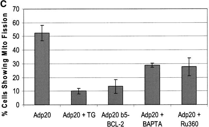
p20-induced mitochondrial fragmentation is mediated by an ER-mitochondria Ca2 + signal. (A) Top, p20 induces a time dependent release of Ca2+ from the ER. H1299 cells were infected with Adp20 in the presence of 50 μM zVAD-fmk and at the indicated times post-infection, cells were loaded with Fura2-AM in Ca2+-free buffer and ER calcium stores were measured as the sudden difference in Fura2 fluorescence recorded after the addition of TG (see Materials and methods). Shown is the mean and SD of five independent experiments. Bottom, elevated [Ca2+]m after Adp20 infection. HeLa cells were treated as in A, except cells were loaded with Rhod2-AM and the [Ca2+]m was estimated as described in the Materials and methods. (B) p20 induces dramatic fragmentation of mitochondria, which is inhibited by predepletion of ER Ca2+ stores with TG. H1299 cells were infected with 50 μM Adp20 + zVAD-fmk in the absence or presence of 50 nm TG for 24 h, and mitochondria were visualized by anti-cyt.c staining. Representative images are shown. (C) Reducing ER Ca2+ stores, chelating cytosolic Ca2+, or preventing mitochondrial Ca2+ uptake inhibits p20-induced fragmentation of mitochondria. As in B, but H1229 cells, or H1299 cells pretreated with 50 nm TG, 2 μM BAPTA-AM, or 20 μM Ru360, or H1299 b5-BCL-2 cells were infected with Adp20 + zVAD-fmk for 24 h and the number of cells showing signs of mitochondrial fragmentation was quantified. Shown is the mean ± SD of five independent experiments.


