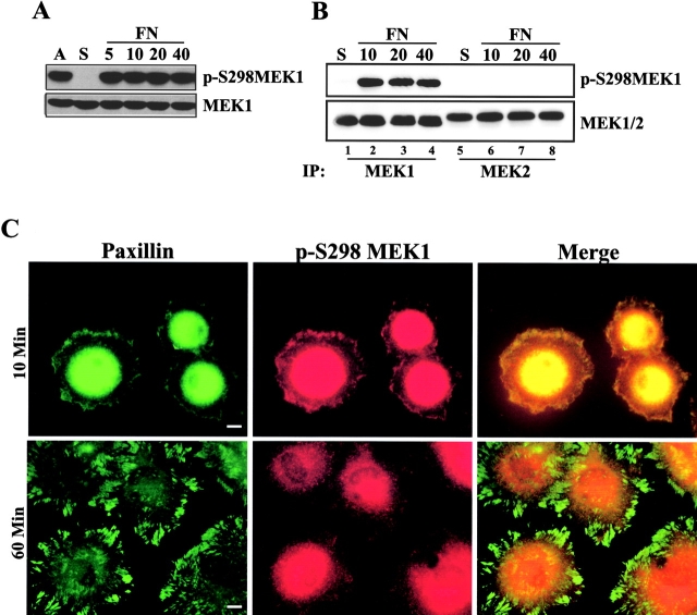
Figure 3.
MEK1 S298 phosphorylation is regulated by cell adhesion. (A) REF52 cells were treated as described in Fig. 1. Whole cell lysates were blotted with antiserum specific for phospho-S298 MEK1 (p-S298MEK1; top) or MEK1 (bottom). (B) REF52 cells were suspended (S) and replated on FN for 10, 20, or 40 min. Anti-MEK1 or anti-MEK2 antiserum was used to immunoprecipitate endogenous proteins, which were subsequently blotted with anti– p-S298MEK1 or anti-MEK1/2. (C) REF52 cells were suspended and plated on FN for 10 min or 1 h before co-staining for p-S298MEK1 (red) and paxillin (green). The intense staining in the center of the cell represents perinuclear staining that was over-exposed to visualize peripheral structures. Bars, 10 μm.

