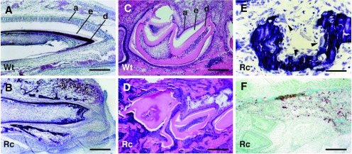
Figure 5.

Tooth histology. (A and B) Sagittal, nondecalcified sections of incisors from a rescued PTHrP-knockout neonate (B) and a wild-type littermate (A) stained with toluidine blue. A well developed ameloblast layer (a) is readily apparent on the labial aspect of the normal tooth (A), but is lacking in the mutant (B), which is crowded by the surrounding alveolar bone. The dark dentin layer (d) is intact but the adjacent enamel (e) is absent from the labial surface of the incisor (the space between the ameloblast layer and the enamel in A is a sectioning artifact). (C and D) Mandibles from 1-week-old wild-type (C) and rescued-knockout mice (D) were decalcified and then sectioned sagittally through the molar crypt. Distortion of the teeth in the rescued-knockout animal caused by progressive impaction is readily apparent. The space between the ameloblast (a) and dentin (d) layers defines the area occupied by the enamel (e) that has been removed through decalcification. (E) Magnification of an nondecalcified sagittal section through the region between the incisor and the first molar of a mandible from a rescued-knockout animal. Osteoclasts (arrowheads) can be seen attached to the surfaces of the alveolar bone. (F) Coronal sections of mandibles from neonatal rescued-knockout animals were stained for tartrate-resistant acid phosphatase activity. Tartrate-resistant acid phosphatase-positive cells (stained brown) are present in the alveolar bone immediately adjacent to the lateral aspect of the molars in a pattern indistinguishable from that found in wild-type littermates (not shown). Wt, wild type; Rc, rescued-knockout. [Bar = 64 μm in (A and B); 32 μm (C, D, and F), and 16 μm (E).]
