Figure 4.

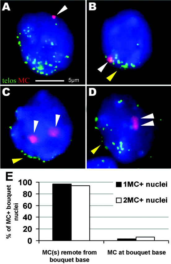
MCs locate remote from the telomere cluster in bouquet nuclei. (A and B) FISH with a (C3TA2)3 PNA telomere probe (green) and an α-satellite DNA MC probe (red; white arrowhead) on monosomic bouquet stage nuclei (1MC+-30; 12 d post partum). (A) A spermatocyte I with one MC located remote from the telomere cluster at the top of the nucleus (the latter faces the observer). (B) Spermatocyte with the MC located among the tightly clustered telomeres at the bouquet base (yellow arrowhead). Focal plane near the nuclear top. (C and D) Bouquet stage nuclei of a 12-d post partum disomic mouse (2MC+-7). (C) Two separate MCs (white arrowheads) located away from the bouquet base (yellow arrowhead). Focal plane at nuclear equator. (D) Spermatocyte I nucleus with relaxed telomere clustering and two paired MCs (white arrowheads) that create a single large MC signal in the nuclear interior below. Focal plane at the nuclear top. The bar in A represents 5 μm and applies to A–D. (E) Distribution frequencies of MCs with respect to the telomere cluster site. Two MCs were generally absent from the telomere cluster region.
