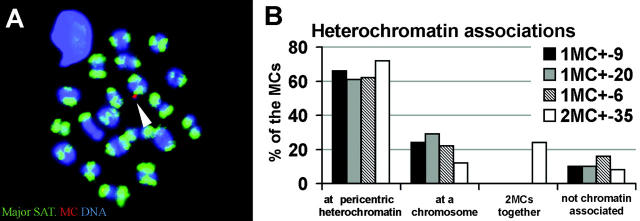
Figure 9.
Heterochromatin associations of MCs in MI. (A) Mouse major satellite DNA FISH (green) and α-satellite DNA MC-FISH (red) on an MI spread shows the MC signal touching the pericentric heterochromatin of a mouse bivalent (arrowhead). (B) Frequencies of MC associations with mouse pericentromeric heterochromatin during the first meiotic metaphase. All mice used, except mouse 2MC+-35, are monosomic MC+ mice. In the MI spermatocytes of mouse 2MC+-35, a total of 25 MCs was scored (nine nuclei with one MC; eight nuclei with two MCs). 8% of these MCs were located together and were also associated with pericentric heterochromatin (number implemented in both bars). Another 8% of the MCs localized next to each other and to the euchromatic part of a mouse chromosome (number implemented in both bars).

