
Figure 1.
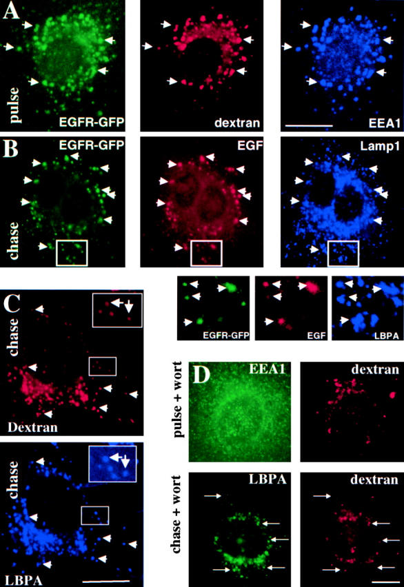
Transport from early to late endosomes after PI3 kinase inhibition. (A) BHK cells expressing EGFR-GFP were preincubated with EGF for 1 h at 4°C and washed; this pretreatment was always used for EGF or EGFR endocytosis. Cells were incubated for 10 min at 37°C with rhodamine-dextran (pulse), labeled with antibodies against the indicated antigens, and analyzed by triple channel fluorescence microscopy. (B) BHK cells overexpressing EGFR-GFP were incubated with EGF-biotin and streptavidin-phycoerythrin for 1 h at 4°C and then for 10 min at 37°C, followed by a 90-min chase in the presence of 0.5 mg/ml leupeptin and analyzed as in described for A; high magnification views of the indicated areas are shown below the micrographs. (C) A pulse of rhodamine dextran was endocytosed as described for A and then chased for 45 min; cells were analyzed as described for A. (D) A pulse of rhodamine dextran was endocytosed as described for A into BHK cells with or without chase (as in C); 100 nM wortmannin (Wort) was present during both the pulse and the chase. Cells were analyzed as described for A. Bars: (A–C) 5 μm; (D) 2.5 μm.