
Figure 1.
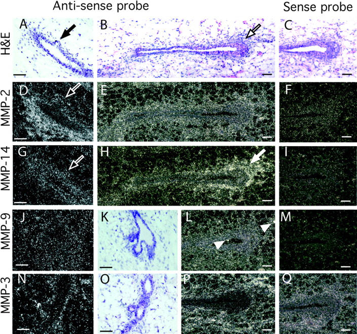
Localization of MMPs-2, -3, -9, and -14 mRNA within the mammary gland. Mammary glands were taken at 50 d old and sectioned. (A–C, K, and O) Hematoxylin and eosin counterstain of mammary gland sections in G–J and N, respectively. Note the initiating lateral branch in A (black arrow) and TEB in B (black outlined arrow). In situ hybridization analysis was performed with the following antisense and sense probes (as indicated): (D–F) MMP-2, (G–I) MMP-14, (J, L, and M) MMP-9, and (N, P, and Q) MMP-3. Note the reduction in MMP-2, but not MMP-14 mRNA, at the initiating lateral branch in the adjacent sections D and G (white outlined arrows); the localization of MMP-14 around the TEB in H (white arrow) and the spots of MMP-9 expression probably localized in macrophages in L (white arrow heads). Bars, 50 μm.