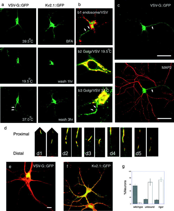
Figure 1.

Post-Golgi transport of axonal and dendrite carriers in hippocampal neurons visualized by CLSM. (a) Sorting of membrane protein–GFP fusion proteins under specific cell manipulation. VSVtsO45-G::GFP localizes in the ER at 39.5°C and moves to the TGN after a 30-min incubation at 19.5°C. Its post-Golgi transport starts when the temperature is shifted to 37°C. Note that VSV-G::GFP carriers are predominantly transported to one neurite (arrows) among the others. Kv2.1::GFP distributes diffusely in the somatodendritic area when expressed overnight in the presence of 1 μM brefeldin A. Kv2.1::GFP accumulates at the Golgi region 1 h after the brefeldin A washout, and starts post-Golgi transport. Note that Kv2.1::GFP was transported to all the dendrites 3 h after wash. (b) Characterization of VSV-G::G(Y)FP probes in neurons. (b1) Neurons were infected with Ad(VSVtsO45-G::GFP) and incubated overnight at 39.5°C, for 30 min at 19.5°C, and for 1 h at 37°C in the presence of 1 mg/ml Texas-red dextran (MW3000). Note that post-Golgi VSV-G carriers in the axon (arrows, green) are not labeled with the endocytotic marker (red). (b2 and b3) Double-label of hippocampal neuron with VSV-G::YFP and Golgi–CFP. At 19.5°C (b2), VSV-G::YFP (b2, green) was colocalized with the Golgi complex marker (b2, red) in the cell body. When the temperature is shifted to 37°C (b3), VSV-G::YFP moves from the Golgi complex area (yellow area at the upper right of b3, due to the overlap of VSV-G::YFP [green] and Golgi–CFP [red]) to the axon (b3, arrows). (c) VSV-G::GFP was dominantly transported from the TGN to the axon (arrow). Neurons were infected with Ad(VSVtsO45-G::GFP) and incubated overnight at 39.5°C, for 30 min at 19.5°C, and for 1 h at 37°C to visualize post-Golgi membrane transport (green). After fixation, the dendrites were stained with the anti-MAP2 antibody (red). Bar, 50 μm. Videos 1 and 2 are available at http://www.jcb.org/cgi/content/full/jcb.200302175/DC1. (d) Post-Golgi axonal carriers of VSV-G::GFP transport various membrane proteins. Axonally transported tubulovesicular organelles were simultaneously double-labeled with CFP- and YFP-tagged proteins, and time-lapse data were collected by sequential activation with 442 and 488 nm lasers by CLSM. (d1) VSV-G::CFP::CFP (red) and GAP-43::YFP (green). (d2) VSV-G::CFP::CFP (red) and β-APP::YFP (green). (d3) VSV-G::YFP (green) and Vamp2::CFP (red). (d4) Vamp2::CFP (red) and GAP-43::YFP (green). (d5) Vamp2::CFP (red) and β-APP::YFP (green). In each set, interval between the right and left panel is 10 s. Slight gaps between the CFP and YFP images along the longitudinal axis of vesicles are due to the time lag (∼0.7 s) between the sequential data acquisition. (e and f) Dominant negative kinesin (S205A,H206A; stained with the H2 antibody; red) inhibits polarized axonal transport of VSV-G::GFP (e, green), whereas it does not inhibit significantly dendrite transport of Kv2.1::GFP (f, green). Bars, 10 μm. (g) Inhibition of polarized axonal transport by dominant negative kinesins. Black bars indicate percentage of neurons with polarized VSV-G::GFP transport, and white bars indicate percentage of neurons exhibiting accumulation of VSV-G::GFP at TGN. Data were collected from four independent cultures.
