Figure 2.
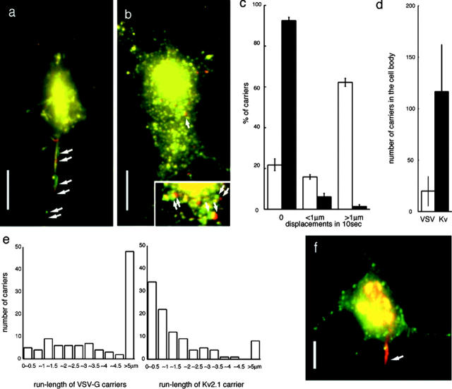
Analysis of axonal and dendrite carrier movement by CAFM. CAFM images of the post-Golgi complex carriers to the axon (a: VSV-G::GFP) and to the dendrites (b: Kv2.1::GFP). Two images at 10-s intervals were merged into one image to show the displacements of individual carriers for 10 s (red images are obtained 10 s later than the green images). Axonal tubulovesicular organelles showed high motility and dominantly transported to the axon (arrows in a indicate three moving carriers). In contrast, post-Golgi carriers for Kv2.1 were vesicular in shape and do not show directional preference in motility (inset in b shows a high magnification of the area indicated by an arrow. Three moving carriers are indicated by arrows in the inset. Note carriers with displacements less than their diameter show overlapping yellow region of two merged images). Corresponding videos are available as Videos 3–5. (c–e) Quantitative analysis of the dynamics of axonal versus dendrite carriers. (c) Percentage of the carriers which shows 0 μm, <1 μm, >1 μm displacements in 10 s in the cell body. Black bar, Kv2.1 carriers; white bar, VSV-G carriers. Data were obtained from three independent cultures. 200 carriers were counted in each culture. (d) Number of the post-Golgi carriers remaining within the cell body. Carriers were counted at 1 h after the start of the post-Golgi transport. Black bar, Kv2.1 carriers; white bar, VSV-G carriers. (e) Histogram of the run-length of individual 100 post-Golgi carriers of Kv2.1 and VSV-G. Carriers, which showed no displacement, were omitted from the histogram. Carriers, which move into neurites and get out of the observing field, were included in the group of >5 μm run-length. (f) Simultaneous double labeling with Kv2.1::YFP (green) and VSV-G::CFP::CFP (red). Note that they colocalize in the Golgi complex area. Kv2.1 carriers distributed in the somatodendritic area, but VSV-G carriers move to the IS (arrow). Bars, 10 μm.

