
Figure 1.
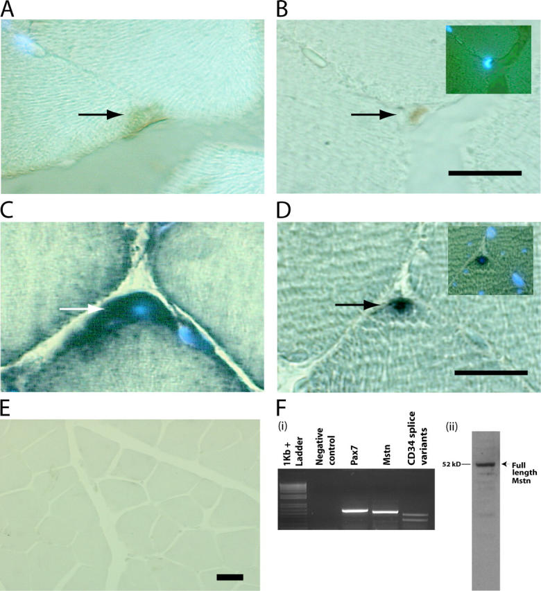
Myostatin is expressed in satellite cells. M. tibialis anterior was serially sectioned and immunostained with antibodies specific for (A) Myostatin and (B) Pax7. The myonuclei were stained with DAPI. The same muscle was used for in situ and probed for Myostatin (C) and Pax7 transcripts (D). Arrows indicate the stained satellite cells, and DAPI-stained myonuclei are also shown in the insets (B and D). (E) Micrograph showing in situ hybridization performed with myostatin antisense probe on muscle sections of myostatin-null mouse. (F, i) Agarose gel electrophoresis of RT-PCR products derived from satellite cell total RNA. Primers specific for 3′ region of myostatin amplify the expected 515-bp product in a combined RT-PCR reaction (Mstn). Amplicons are not detected in the absence of template (Negative control). Pax7 was amplified with primers designed to produce a 571-bp product (Pax7), and CD34 splice variants were also PCR amplified from the same RT reaction (CD34 splice variants). 1-kb plus DNA ladder is shown. (F, ii) Western blot showing the presence of the full-length 52-kD Myostatin protein in satellite cell protein extract. Bars, 10 μm.