
Figure 4.
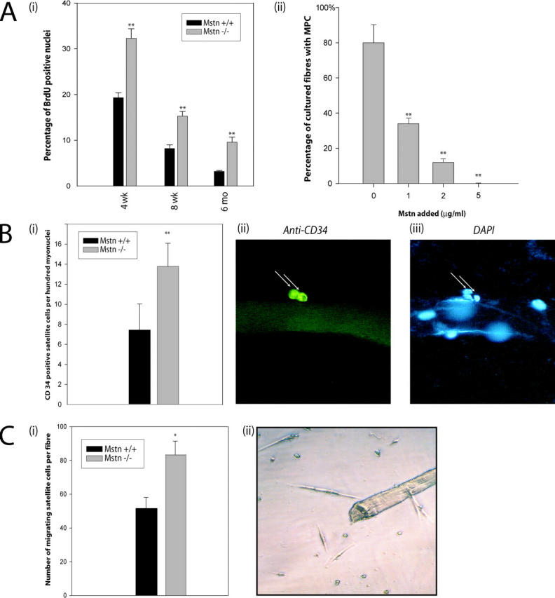
Lack of Myostatin increases the activation of satellite cells on myofibers. (A, i) In vivo quantification of activated satellite cells in the muscle of wild-type and myostatin-null mice. Activated satellite cells were labeled with BrdU in wild-type or myostatin-null (Mstn −/−) mice of 4 wk, 8 wk, or 6 mo and were isolated using Percoll gradient. 5,000–10,000 satellite cells were immunostained for BrdU, and percentages of nuclei that were positive for BrdU labeling are shown. **, P < 0.01 (as compared with wild type). At least a total of 1,000 cells were counted in each of three replicates. The data provided are an average of three animals each. (A, ii) Myostatin inhibits the migration of satellite cells from fibers. Single muscle fibers (n = 32) were isolated from the muscle and incubated in media conducive to the migration of myogenic precursor cells. The addition of Myostatin in increasing concentrations decreases the percentage of fibers with migrated satellite cells (**, P < 0.01). (B, i) The number of satellite cells (CD34 positive) per 100 myonuclei in wild-type and myostatin-null (Mstn−/−) myofibers is shown. **, P < 0.01 (as compared with wild type). More than 1,000 nuclei were counted in each of three replicates. The data presented are an average of three animals each. (B, ii) Micrograph showing typical CD34-immunostained satellite cell and (iii) myonuclei were visualized by counterstaining with DAPI. (C, i) An increased number of satellite cells migrate from myostatin-null fibers. Quantitative analysis demonstrates that an increased number of myogenic progenitor cells migrates from single myofibers isolated from myostatin-null mice as compared with wild-type myofibers (*, P < 0.05). (C, ii) An example of satellite cells surrounding an isolated myofiber after 72 h.