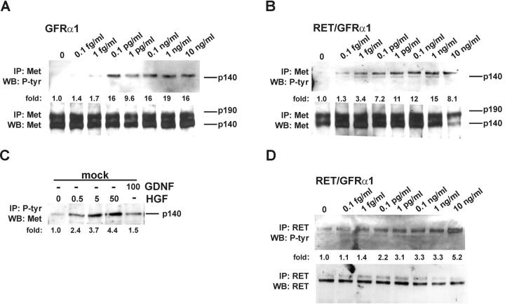
Figure 4.
GDNF induces phosphorylation of Met. (A and B) Dose-dependent phosphorylation of Met by GDNF in GFRα1- and Ret/GFRα1-expressing MDCK cells. Met was activated in 15 min after GDNF application. The bottom panels show the reprobing of the same filter with anti-Met antibodies. The numbers below the lanes indicate the fold of induction of Met tyrosine kinase. (C) Phosphorylation of Met in mock-transfected MDCK cells. Concentrations of GDNF and HGF are given in ng/ml. 30 μg of total proteins were incubated with 10 μl of immobilized phosphotyrosine mAbs, and immunocomplexes were washed and analyzed as described in Materials and methods. (D) Dose-dependent activation of Ret by GDNF in Ret/GFRα1-expressing MDCK cells. The bottom panel shows the reprobing of the same filter with anti-Ret antibodies. The numbers below the lanes indicate the fold of induction of Ret tyrosine kinase. IP, immunoprecipitation; WB, Western blotting; P-tyr, phosphotyrosine. The results are representative of three independent experiments.

