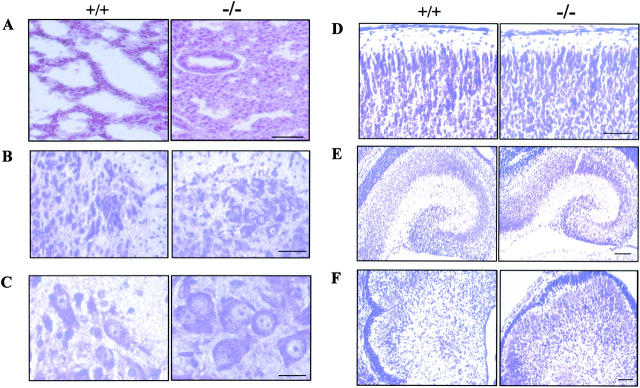
Figure 2.
Histology of KIF5A null mutant mice. (A) Lung histology of KIF5A null mutant. 7-μm lung paraffin sections from KIF5A null (KIF5Anull/KIF5Anull) and control (KIF5AWT/KIF5AWT) littermates were stained with hematoxylin and eosin. Note that the mutant lung was not well expanded. Bar, 50 μm. (B–F) Histology of KIF5A null mutant nervous tissues. Paraffin sections from spinal cord (B and C), cortex (D), hippocampus (E), and cerebellum (F) were stained with cresyl violet. Note that no obvious differences were observed between KIF5A null mutant and control littermates except that the cell bodies of the motor neurons were larger in the mutant spinal cord. Bars: (B, D, and F) 50 μm; (C) 20 μm; (E) 100 μm.

