
Figure 3.

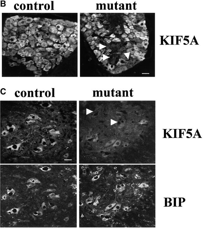


Gross analysis of KIF5A conditional mutant mice (KIF5A nul l/KIF5Aflox; Cresynapsin). (A) Western blot analysis to measure KIF5A protein levels in the brains of KIF5Anull/KIF5Aflox; Cresynapsin mice. Equal amounts of brain homogenate from 3-wk-old mutant and control (KIF5Aflox/KIF5AWT) littermates were loaded; actin was used as a loading control. Each marked number represents the ratio between the mutant band and control band after normalizing with the actin band. (B and C) KIF5A excision by Cresynapsin transgene in DRG (B) and spinal cord motor neurons (C) of KIF5Anull/KIF5Aflox; Cresynapsin mutant. Tissue sections from ∼3-wk-old mutant and control littermates were stained with KIF5A-specific antibody. Spinal cord sections were also double stained with an anti-BIP antibody to visualize the motor neuron cell bodies. Note the decreased or lack of KIF5A staining in some neurons (arrowheads). Bar, 100 μm. (D) A comparison of body weight (mean ± SD) among 3-wk-old littermates with different genotypes. Note the obvious low body weight in the KIF5Anull/KIF5Aflox; Cresynapsin mutant group, n = 4 for each group. *, P < 0.01. (E) Most KIF5Anull/KIF5Aflox; Cresynapsin mutant mice died around 3 wk of age. Postnatal survival curve of a group of mice (142 total, 113 control and 29 mutant) is shown here. The rate of survival of the different genotypes was plotted against age. (F) Abnormal hind limb posture in an older KIF5Anull/KIF5Aflox; Cresynapsin mutant mouse. Two 7.5-mo-old littermates (control and KIF5Anull/KIF5Aflox) are shown.