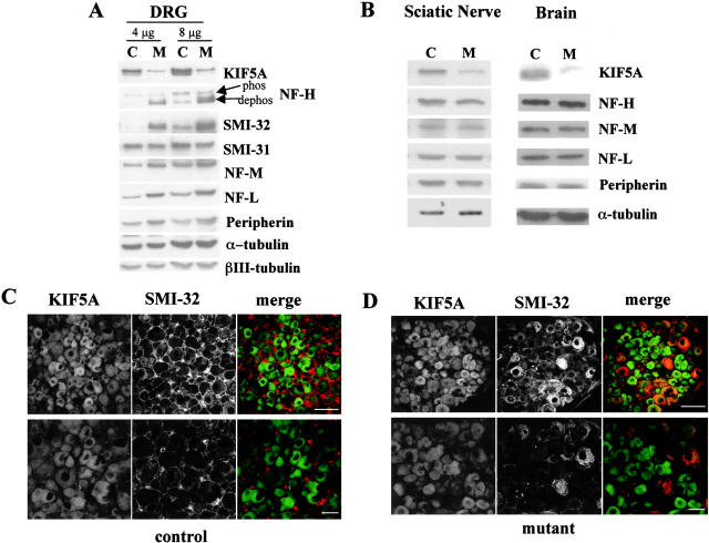
Figure 6.
Accumulation of NF proteins in DRG sensory neuron cell bodies of KIF5A null /KIF5Aflox; Cresynapsin mutant mice. (A) Western blot analyses of DRGs from the first cohort of 3-wk-old KIF5Anull/KIF5Aflox; Cresynapsin mutant mice. In the litter shown, control DRGs (C) were pooled from one II/+ and one Cre/+ littermates; mutant DRGs (M) were pooled from two mutant littermates. Note the obvious increase in dephosphorylated NF-H (revealed by the lower band labeled by NF-H, a polyclonal antibody against the COOH terminus of NF-H, and by antibody SMI-32) as well as the increase in NF-M, NF-L, and peripherin. (B) Western blot of brain and sciatic nerve from 3-wk-old KIF5Anull/KIF5Aflox; Cresynapsin mutant mice. 20 μg of proteins of mutant and control (II/+) littermates was loaded in each lane. Note the clear reduction of KIF5A protein in the mutant. The levels of NFs and peripherin were not significantly changed in the mutant brain and sciatic nerve. (C and D) Immunostaining of DRG sensory neurons of 7.5-mo-old KIF5Anull/KIF5Aflox; Cresynapsin mutant mice. Double staining with KIF5A (green) and SMI-32 (recognizes dephosphorylated NF-H, red) was performed on a mutant and a control littermate. (C) Control DRG staining. Bottom panel, higher magnification. (D) Mutant DRG staining, higher magnification in the lower panel. Note the apparent intense SMI-32 staining in some DRG neurons. Bars, 200 μm.

