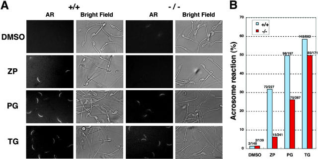
Figure 2.
Requirement of PLCδ4 in ZP- and progesterone-induced acrosome reaction. (A) Fluorescence and phase images of sperm treated with several acrosome reaction–inducing reagents. PLCδ4+/+ (+/+) and PLCδ4−/− (−/−) sperm capacitated for 1 h were treated with 1.0% DMSO, 3 Zp/μl solubilized mouse ZP (ZP), 100 μM progesterone (PG), or 5 μM thapsigargin (TG) for 15 min at 32°C in the presence of 2 μM Alexa Fluor®594–conjugated SBTI to monitor the occurrence of the acrosome reaction. The same microscopic fields are shown for acrosome reaction (AR) and bright field images. Bar, 10 μm. (B) Percent of sperm that underwent the acrosome reaction. The numbers above each column represent the number of sperm examined. Data were obtained from total amount of sperm by four independent experiments for progesterone and ZP and three independent experiments for thapsigargin.

