
Figure 4.
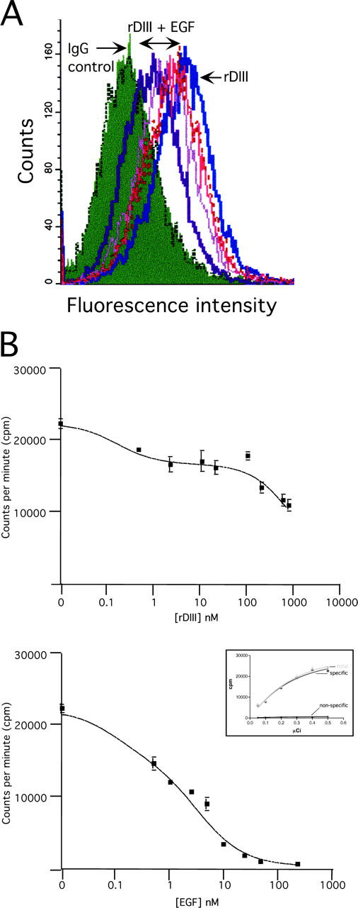
Competitive binding of rDIII and EGF. (A) Flow cytometry. MDA-MB-231 cells were incubated with 2.00 μM rDIII, in the presence of increasing concentrations of EGF (0.45, 0.85, 1.25, and 2.00 μM). rDIII binding to the cell surface was detected with anti-His tag and Alexa® 488 antibodies. The fluorescence signal for rDIII gradually decreases with increasing EGF concentrations. (B) Displacement of cell surface-bound I125-EGF by rDIII. MDA-MB-231 cells were incubated with I0.5 nM 125-EGF and increasing concentrations of cold rDIII (top) or EGF (bottom). The 0.5 nM (≈0.15 μCi) working concentration of I125-EGF was determined by calculating the specific binding of I125EGF (“specific”) based on total and nonspecific binding of I125-EGF to MDA-MB-231 cells (inset in bottom panel). Cells were incubated with increasing concentrations of I125-EGF in the absence (total binding; “total”) or presence of an excess amount (330 nM) of unlabeled EGF (nonspecific binding; “nonspecific”).