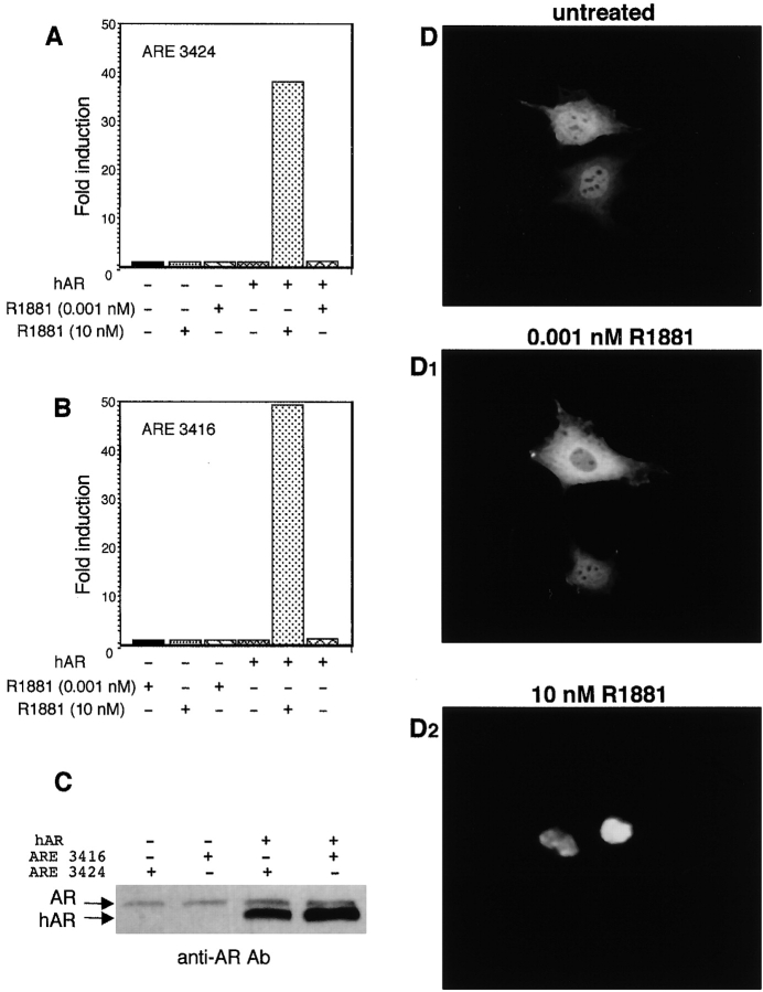
Figure 6.
Androgen-stimulated gene transcription in NIH3T3 cells before and after overexpression of hAR and hAR intracellular localization. NIH3T3 cells were transfected with either ARE 3424 (A) or 3416 (B) constructs with or without hAR-expressing plasmid. Cells were left unstimulated or stimulated for 18 h with the indicated concentrations of the androgen R1881. The luciferase activity was assayed, normalized using β-gal as an internal control, and expressed as fold induction. The same lysates were used for Western blot analysis with C-19 anti-AR Ab (C). (D and D2) NIH3T3 cells were transfected with hAR-expressing plasmid, and then made quiescent. Cells were left unstimulated (D) or stimulated for 1 h with either 0.001 nM (D1) or 10 nM R1881 (D2). Fixed cells on coverslips were permeabilized as described in Materials and methods, and hAR was visualized by immunofluorescence using the rabbit polyclonal anti-AR (N-20) antibody.

