
Figure 3.
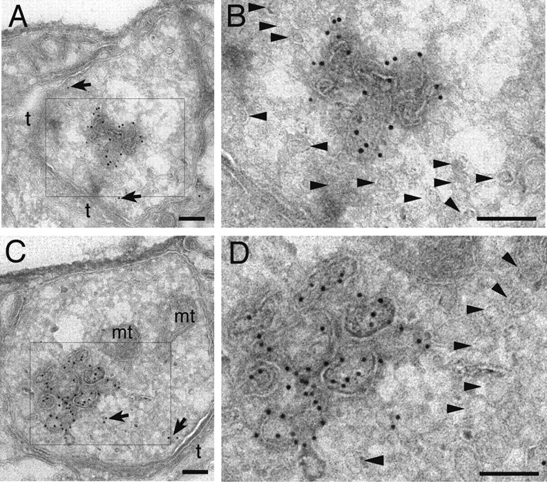
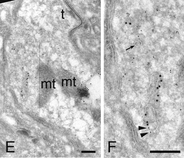

Cryoimmuno-EM of the GFP-2xFYVE endosome. (A–D) Cryoimmuno-electron micrographs showing two Drosophila presynaptic terminals (A/B and C/D), where GFP-2xFYVE is labeled by 10-nm gold particles (anti-GFP antibody). (B and D) High magnifications of the boxes in A and C, respectively. We found cisternal structures of around 150 nm associated to a more electron-dense region within the terminal. The darker regions allow a better contrast for visualization of the membrane (which appear lighter in cryosections) associated to the endosomes, compared with the vesicles with a diameter of ∼35 or 70 nm (see also Fig. 7). Vesicles are, however, occasionally observed (arrowheads). Only few gold particles (7.8 ± 1.3%, n = 5 sections) are associated to the vesicles (arrows). Cryoimmuno-electron micrographs showing localization of GFP-2xFYVE (10-nm gold particles) and endogenous CSP (5 nm gold; E and F) or endogenous Rab5 (5 nm gold; G and H). (F and H) High magnifications of the boxes in E and G, respectively. (E and F) CSP appears throughout the bouton area associated to the pool of vesicles, whereas GFP-2xFYVE is largely restricted to the cisternal endosomal compartments. Although not many vesicles are distinguishable (F, arrow), their presence is revealed by staining of SV integral membrane protein CSP. Few 5-nm gold particles labeling CSP could also be observed in the cisternal structures (F, arrowheads). Rab5 appears in the cisternal structures, (H, arrowheads) as well as in other regions corresponding to vesicles or cytosol (H, arrows). Note that immunodetection is highly specific, because neither 10-nm (GFP-2xFYVE) or 5-nm gold particles (CSP and Rab5) were very rarely detected in the postsynaptic subsynaptic reticulum or the mitochondria in the cryosections (<1% of the gold particles; A–H; unpublished data). t, T-bar or electron-dense regions indicating active zones; mt, mitochondria. Bars: (A–D) 150 nm; (E–H) 200 nm.