
Figure 1.
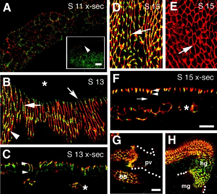
Gli is expressed in the epidermis and it is localized to the tricellular corners of abutting epithelial cells. (A–H) Whole mount embryos of different developmental stages are shown double stained for Gli (green), and either the (A–F) pSJ protein Nrx or (G and H, red) Cora. The overlap of Gli and Nrx, or Cora, is yellow. Images are of wild-type embryos, with the exception of E, which is a Gli AE2Δ 45-null mutant. (A) High magnification view of the epidermis (cross section view) of a stage 11 embryo taken at the position of the arrowhead in the low-magnification inset. At this stage, the distribution of Gli is quite uniform around the surface of the epithelial cells and similar to that of Nrx. (B) En face view of a stage 13 embryo undergoing dorsal closure. The localization profile for Gli is variable. At the leading edge (concave arrow), Gli diffusely labels epithelial cell membranes as they make contact with the underlying amnioserosa (asterisk). Epithelial cells in more ventral positions, have Gli concentrated in patches around their circumferences (solid arrow), or localized to tricellular corners (arrowhead). Gli distribution is distinct, although overlapping, with that of Nrx. The amnioserosa lacks both Gli and Nrx expression (asterisk). (C) Cross section of a stage 13 embryo. Gli expression is restricted to the lateral membrane of epithelial cells (region between arrowheads), and is concentrated in patches. Gli staining in the tracheae underlying the epidermis is also evident (asterisk). (D) En face view of epidermis of a stage 15 embryo. Gli remains concentrated at tricellular junctions (arrow). (E) En face view of a stage 15 Gli AE2 Δ45-null mutant, double stained for Gli and Nrx. In the absence of Gli protein, the Gli mAb does not stain the tricellular corners of the epidermis (arrow) and, thus, it is specific to a Gli epitope. (F) Cross section of the epidermis of a stage 15 embryo. Gli localization is restricted to the apical portion of the lateral membrane domain and lies within the Nrx-positive SJ domain (arrowheads). Gli expression does not extend to the bottom of epithelial cells (arrow). Gli is only sporadically seen in the lateral membrane of epithelium because the plane of section only occasionally transects a tricellular corner. Gli expression in tracheae persists (asterisk). (G) Gli expression is evident in the esophagus (es) as is Cora. Both proteins are absent from the outer layer of the proventriculus (pv, dotted line). (H) Gli and Cora are expressed in the hindgut (hg), but absent from midgut (mg). Bars: (A, inset) 50 μm; (F and G) 10 μm.