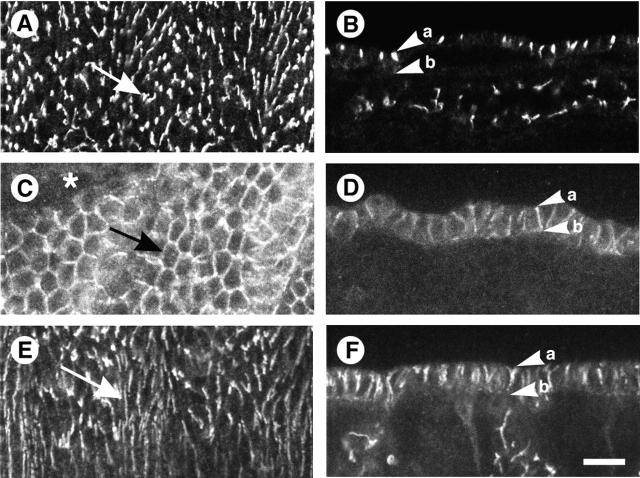
Figure 3.
Gli localization at epithelial tricellular corners is dependent on Nrx. All embryos are stained for Gli and are at stage 15 of development. (A and B) Wild-type embryos. (C–F) Nrx 46 homozygous mutant embryos. (A) Gli localizes to the tricellular corners (arrow) of epithelial cells (en face view). (B) Gli, at tricellular corners, is restricted to the apical half of the lateral membrane domain (cross section view). The apical (a) and basal (b) surfaces of an epithelial cell are marked. (C and D) Severe Nrx 46 homozygous mutant embryo. (C) Gli fails to localizes to the tricellular corners of epithelial cells (arrow) and is distributed around the circumference (en face view). In this late stage embryo, dorsal closure has failed (asterisk). (D) Cross section view of the epidermis in C. Rather than being restricted to the apical half of the lateral membrane, Gli extends to the basal surface. (E and F) Nrx 46 homozygous mutant embryo with a weak dorsal closure phenotype. (E) En face view of the epidermis. In these mutants, the epidermis appears more wild type, but Gli does not localize to tricellular corners (arrow). (F) Cross section view of the epidermis in E. As in D, Gli is not concentrated to the pSJ domain at tricellular corners, but diffuses basally. Bar, 10 μm.

