
Figure 4.
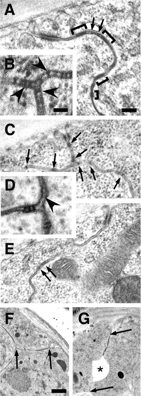
TEM of stage 17 Gli mutant embryos reveals that pSJ septae are not tightly arrayed. (A,B, and F) Wild-type epidermis. (C–E and G) Gli AE2Δ 45 mutant epidermis. (A) pSJs typically contain clusters of septae (brackets) interspersed with single or pairs of septa (arrows). (B) At tricellular junctions, pSJ septae are present at all bicellular contacts (arrowheads). (C and E) In Gli mutants, single or pairs of pSJ septa predominate (arrows), and few clusters are evident. (D) pSJ septa are not present at all bicellular contacts of tricellular junctions in Gli mutants (arrowhead). (F and G) Embryos prepared for TEM analysis with high pressure freezing. (F) In wild type, neighboring epidermal cells contact each other over the length of the lateral membrane (region between arrows). (G) In Gli mutants, large regions of delamination (asterisk) are observed in the lateral membrane domain (region between arrows) of >30% of contacting cells, as opposed to 5% in wild type. Bars: (B and D) 50 nm; (F and G) 400 nm; (A, C, and E) 100 nm.