
Figure 1.
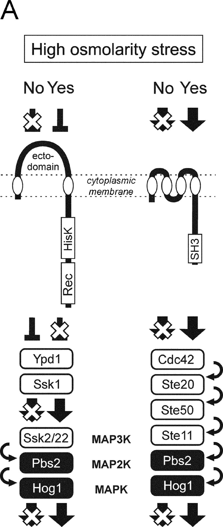
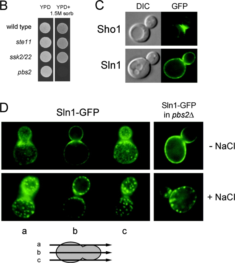
Sln1 and Sho1 have distinct cellular distributions. (A) Architecture of the SLN1 and SHO1 branches of the HOG pathway. (B) Either the SLN1 or SHO1 branch is sufficient to survive on high osmolarity. The wild-type, ste11Δ, ssk2Δ ssk22Δ, or pbs2Δ mutant cells were spotted on low (YPD) and high (YPD + 1.5 M sorbitol) osmolarity media plates, and were grown for 2 d at 30°C. (C) Sln1 has a nearly uniform cytoplasmic membrane distribution. The localization of the Sln1–GFP and the Sho1–GFP fusion proteins was analyzed by fluorescent microscopy in unstressed cells. GFP, fluorescence images; DIC, differential interference contrast images. (D) Hyperosmotic stress induces a clustering of Sln1. Wild-type or pbs2Δ mutant cells expressing Sln1–GFP were observed by fluorescent microscopy before and after (5 min) addition of 0.4 M NaCl.