
Figure 1.
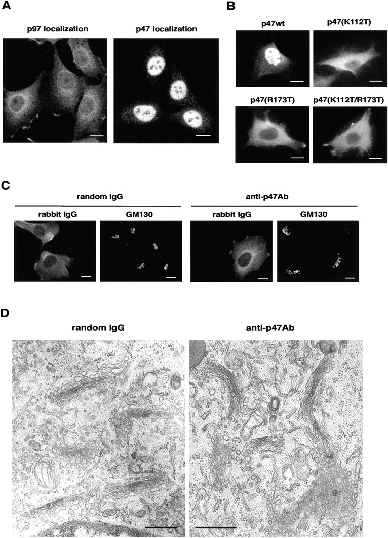
p47 is mainly localized to a nucleus at interphase. (A) The distribution of p47 and p97. NRK cells were fixed with PFA, permeabilized with Triton X-100, stained with polyclonal antibodies to p97 and p47, and observed by confocal microscopy. Bar, 10 μm. (B) HA-tagged p47 and its mutants were expressed in NRK cells, stained with antibodies to HA tag, and observed by conventional fluorescence microscopy. Bar, 10 μm. (C) NRK cells were microinjected in their cytoplasmic regions with either anti-p47 antibodies (8 g/l) or random IgG (8 g/l) and fixed after 2 h incubation. Injected antibodies and Golgi were stained by anti–rabbit antibodies and monoclonal anti-GM130 antibodies. Bar, 10 μm. (D) Representative EM images of Golgi in the cells microinjected with either anti-p47 antibodies (16 g/l) or random IgG (16 g/l). Bars, 0.5 μm.